NIRS-EEG lab
Introduction
With this measuring instrument it is possible to measure brain activity with a high temporal resolution (EEG) in combination with a reasonable (sub-cm) spatial resolution (NIRS). These measurements are non-invasive, not harmful or damaging for the subject and completely silent. Almost anyone can undergo the NIRS-EEGmeasurement without stress. During the measurements, we can offer visual stimuli (varying from simple lights to movie material) as well as aural stimuli (varying from very simple tones to complex sounds such as speech or music) and study the reactions of the brain in these different conditions. Additionally, we provide the data-analysis and presentation.
The measurements can especially be used for patients with an implant, such as a cochlear implant, hearing aid instruments, or a bone conduction stimulator. Companies producing such implants could gain more insight into the manner in which their products steer brain activity. Audiological centers can use this technique for the same reasons.
Scientific supervisor Marc van Wanrooij
Radboud Research Facilities ›Facilities ›Neurology and Motion ›NIRS-EEG
Experiment information
- If you wanna book the NIRS-EEG Lab, please use Qreserve .
- Please, take notes in the lab journal about experiments that have been run
- Use protocols for your study/experiments/pilots
Prepared NIRS experiments
- Basic finger tapping experiment (blocked)
- Mapping experiments (blocked)
- Auditory oddball experiment (tones, event-related)
Prepared EEG experiments
- Frequency oddball
- Spatial oddball
Basic NIRS analysis
Besides Marc's scripts for all steps during NIRS data analysis, you can start using the fieldtrip toolbox http://www.fieldtriptoolbox.org/development/nirs. Here is a general pipeline of what you may wanna do with your NIRS data.
- read raw data (.oxy3) recorded with the oxymon
- read trigger information from AD channels
- define your trial (pre- and poststimulus interval, event code)
- apply a filter
- segment continuous data into trials (epoching)
- convert data, i.e. changes in optical density to concentration changes
- average data per condition/event
- perform baseline correction
- plot single channels or a whole grid
- More things to do/program: do a reference channel subtraction, plot a topomap
Acquired data using the Polybench Software (TMSI) can be read into Matlab using tms_read(Filename.Poly5) provided by TMSI. Further analysis could for example be run with Fieldtrip (http://www.fieldtriptoolbox.org) or EEGLab (http://sccn.ucsd.edu/eeglab/).
- reformat raw data structure to be compatible with the output of fieldtrip preprocessing
- readout trigger/event information from the trigger channel
- define your trial (pre- and poststimulus interval, event code)
- segment continuous data into trials (epoching)
- detect and remove artifacts
- filtering
- average data per condition/event
- correct baseline
- prepare channel layout once depending on the used electrode cap (for correct plotting)
- plot single channels, all channels, or topographic distribution (for a specified time interval)
Technical information
TDT
- 1 RZ6
- 1 RP2
- 2 RA16 (medusa)


EEG
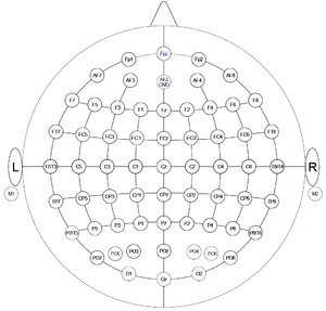
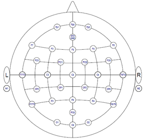
- 32 channel water-based electrode system (TMSI, Mobita), including headcap(s)
- 72 channel REFA, including headcaps in 3 different sizes (TMSI)
- 16 channel REFA (TMSI)
- ring electrodes including separate caps for combined measurements with NIRS
- TMSI Polybench recording software
NIRS
- 2 x 24 channel system (Oxymon, Artinis) each consisting of 8 transmitters and 4 receivers (splitted fibers)
- sampling rate up to 250 Hz
- 8 AD channels (according to Artinis, they only sample at the rate of all other channels. Care needs to be taken, if sampling at low rates. Present your triggers for longer durations then)
- Oxysoft recording software